Inflamed Coronary Arteries: How can medical imaging help to asses a risk of heart attack?
The heart keeps our blood in motion and supplies the cardiovascular system and thus our organs with nutrients. When blood vessels are narrowed because of sediments, known as plaque, the blood circulation is affected. This disease of the coronary vessels is called atherosclerosis which is the most common cause of heart attack. A patient’s heart attack risk is not determined by how narrow a coronary artery is, but by the degree of artery wall inflammation as inflammatory processes in the vessel walls are responsible for the rupture of the vessel wall. Subsequently, a blood clot develops that blocks the vessel and leads to heart attack.
In order to visualise and judge local and temporal dimensions of such inflammations immunologist Dr. Thomas Vogl, chemist Dr. Andreas Faust and nuclear medicine specialist Dr. Sven Hermann work together to develop tracers for molecular imaging. Smokers as well as people with high blood pressure, diabetes, elevated blood lipid levels or other known risk factors, suffer heart attacks more frequently than others. For people belonging to one of these high-risk groups these new imaging methods are aimed at determining whether, in their particular case, the disease is actually present and thereby their individual risk of heart attack is high. For patients already diagnosed with atherosclerosis the new methods will allow checking whether there is an acute risk of heart attack.
Photos

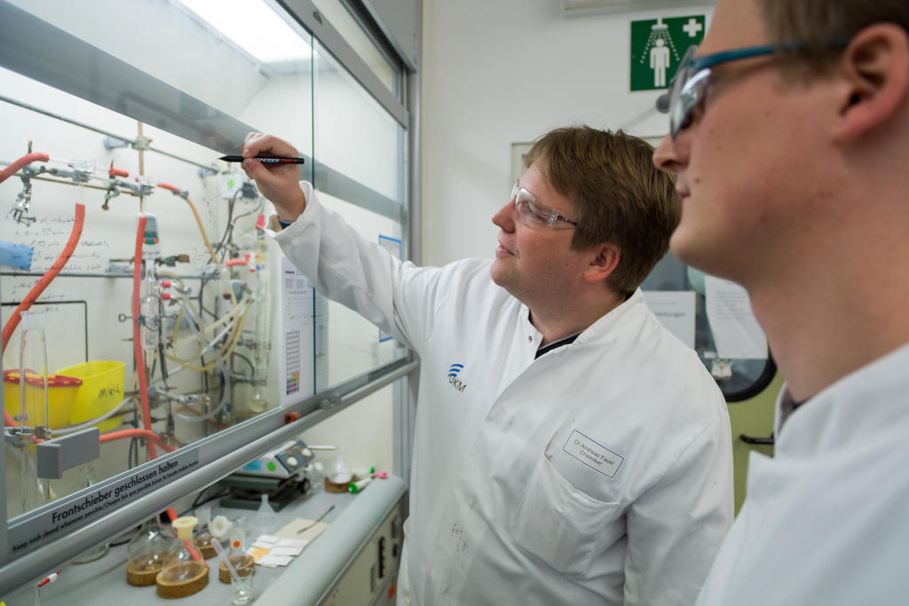
As imaging technique, molecular imaging is used. Dr. Andreas Faust (left) develops new tracer substances in the radiochemistry lab.© SFB 656 - Peter Leßmann 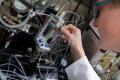
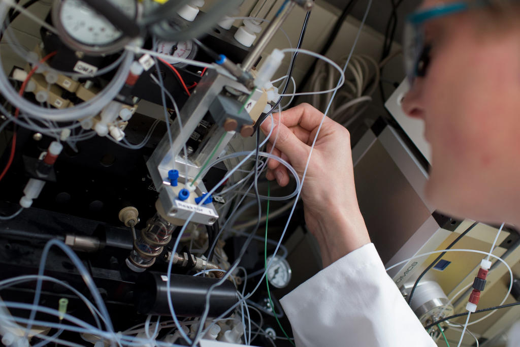
To be able to visualise the tracer substance in the organism, Felix Busch labels it with a weak dose of a radioactive substance.© SFB 656 - Peter Leßmann 
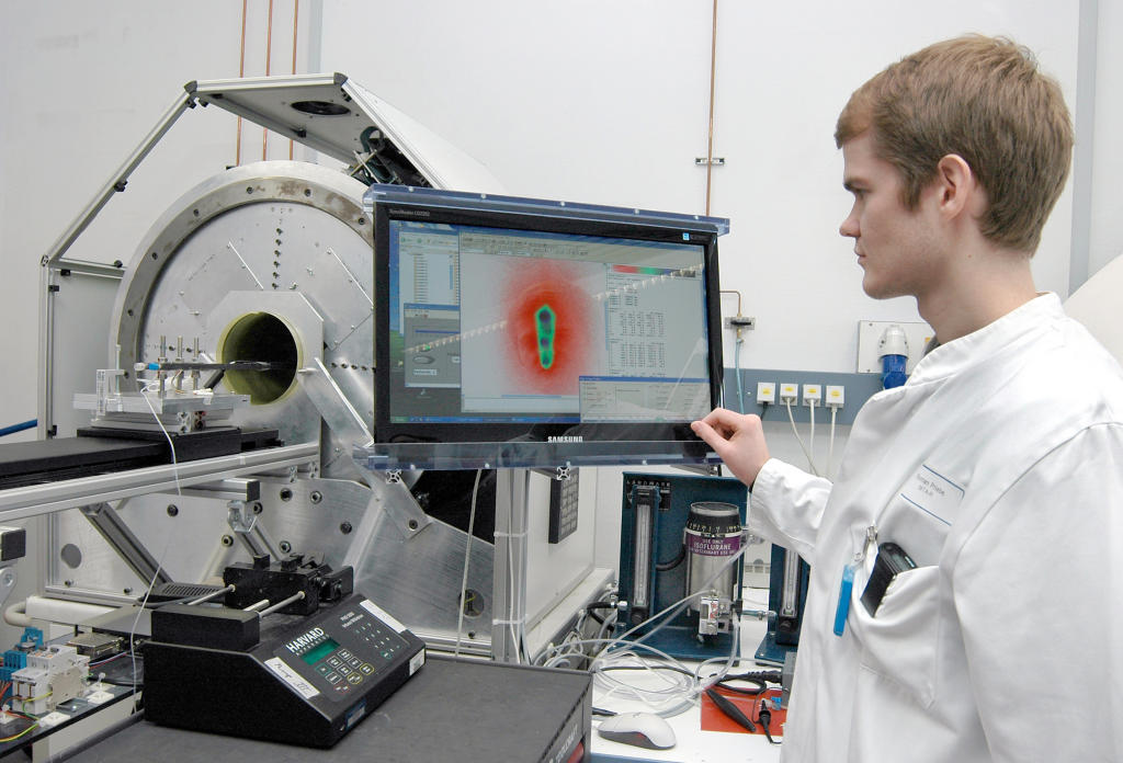
The tracer substance is injected into the organism where it binds to its target molecules in the inflamed vessel wall. With positron emission tomography, the radiation emitted by the tracer can be detected.© SFB 656 - Elisabeth Deiters-Keul 
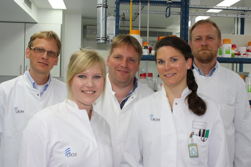
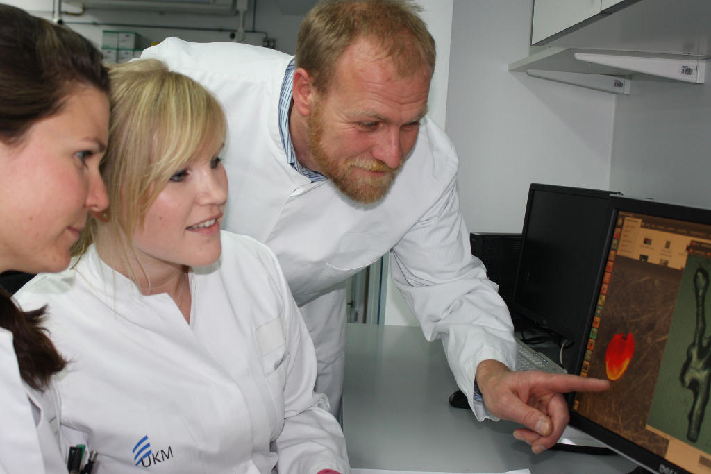
The tracer substance developed by the team led by Dr. Sven Hermann, Dr. Andreas Faust and Dr. Thomas Vogl (left to right), could help to assess the risk of an inflamed vessel wall to rupture in future. This would help in the prevention of heart attack. © SFB 656 - Frieda Berg