Hybrid Imaging with PET/MRI – Innovative Device Combines Two Methods of Imaging the Body
To diagnose cancer or other diseases and to analyse the success of a therapy, it is very helpful to have a look into the body. To do so, physicians make use of several imaging techniques – these have become an integral part of the clinical workflow. Every single method has different assets.
Just recently the University Hospital of Münster (UKM) has brought into operation the hybrid device PET/MRI which combines positron emission tomography (PET) and magnetic resonance imaging (MRI). Overall there are only six PET/MRIs in the whole of Germany. Before installing the hybrid scanner suitable rooms had to be constructed: a control area that shields the magnetic waves coming from the MRI. The advantages of PET/MRI are explained in our podcast by Prof. Michael Schäfers (Director of the Department of Nuclear Medicine at the University Hospital Münster, Co-Coordinator of the Cells-in-Motion Cluster of Excellence and Spokesman of CRC 656). The combination of both imaging techniques brings two different medical departments closer together: the nuclear physicians around Professor Michael Schäfers and the radiologists around Professor Walter Heindel of the University Hospital Münster now take a look at the pictures of the PET/MRI together. As a team, they want to make use of the full potential of PET/MRI.
Photos
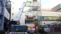
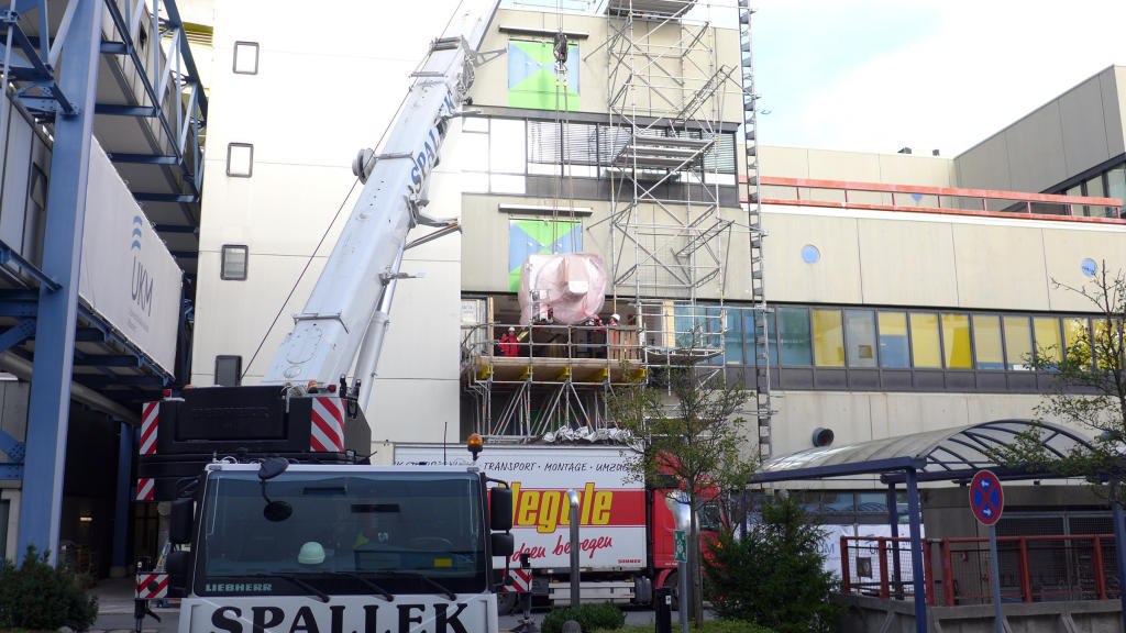
The installation of the new scanner required extensive renovations. To get it into the university hospital the outer wall had to be breached.© Thomas Isokeit 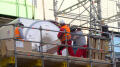
This procedure was the only way to get the device into the Department of Nuclear Medicine where a necessary control area for radiation protection already existed.© Thomas Isokeit 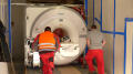
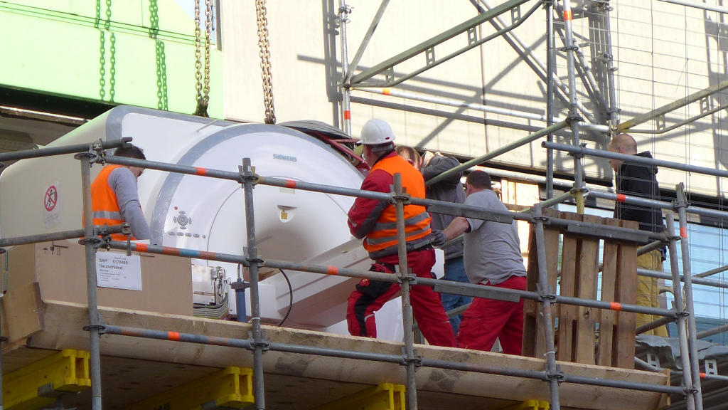
The PET/MRI on the way to its new home. Because of the MRI-component the rooms also had to be shielded against magnetic waves.© Thomas Isokeit 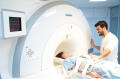
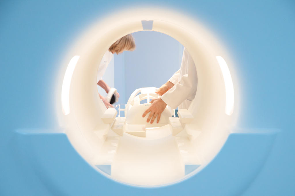
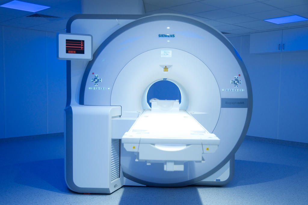
The hybrid PET/MRI device at the University Hospital Münster is already in use to diagnose patients.© Jahn Müller 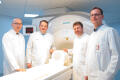
For a combined device you need interdisciplinary expertise: (f.l.t.r.) Prof. Dr. Walter Heindel (Radiology), Prof. Dr. Michael Schäfers (Nuclear Medicine), Dr. Thomas Allkemper (Radiology) und Dr. Lars Stegger (Nuclear Medicine).© Jahn Müller 
To make use of the full potential of PET/MRI the scientists in Münster train physicians to work with both components. In addition they investigate in optimizing the image quality of the scans.© Jahn Müller